Introducing
GAIA
Super resolution for live cell imaging
GAIA
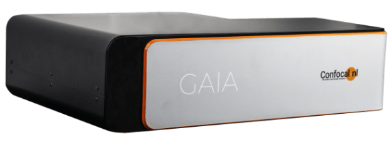
Thanks to its patented REscanning confocal technology, GAIA Point REscan is a super resolution microscope enabling deep live cell imaging beyond the diffraction limit using only nanowatts of power. Our Point REscan is available in two versions, streamlined GAIA α and flagship GAIA λ.
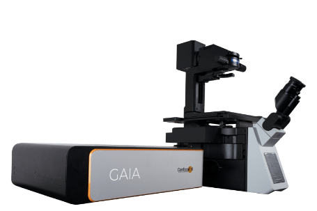
Image wide in super resolution
REvolutionary REdesign of REscan technology in GAIA provides more than double the field of view in the sample plane compared to its predecessor. As a consequence, GAIA enables super resolution imaging over a large FoV and using a wide range of objectives (30X -100X). For the best images high NA objectives are a must.
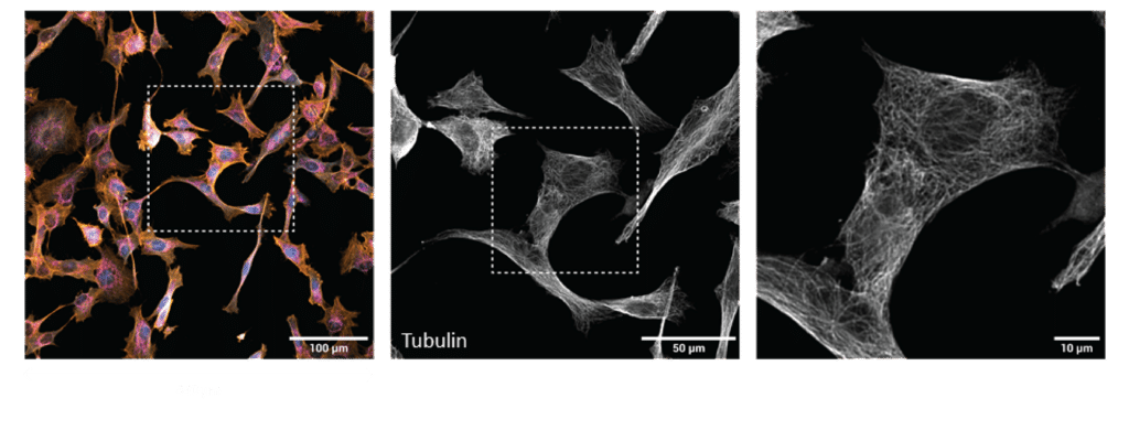
Fibroblasts (30X 1.05NA): Actin 640nm (yellow), Tubulin 561nm (white), Mitochondria 488nm (magenta), Nucleus 405nm (cyan).
Image deep in super resolution
Combination of low laser power requirements, high sensitivity of the detector and a novel optical design present in GAIA Point REscan, enables super resolution beyond 500μm of depth. This makes GAIA Point REscan confocal a perfect solution for live cell super resolution imaging of even thicker specimens like cleared tissue, whole zebrafish embryos, organoids and spheroids.
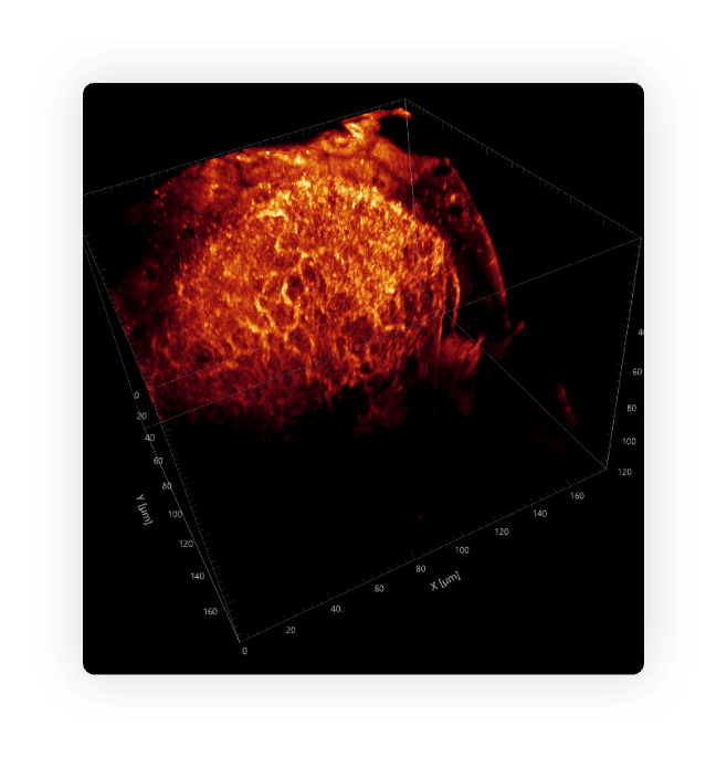
Optic tectum of Zebrafish. Image courtesy of Bram Willems (Brinks Lab, TU Delft).
Explore the benefits
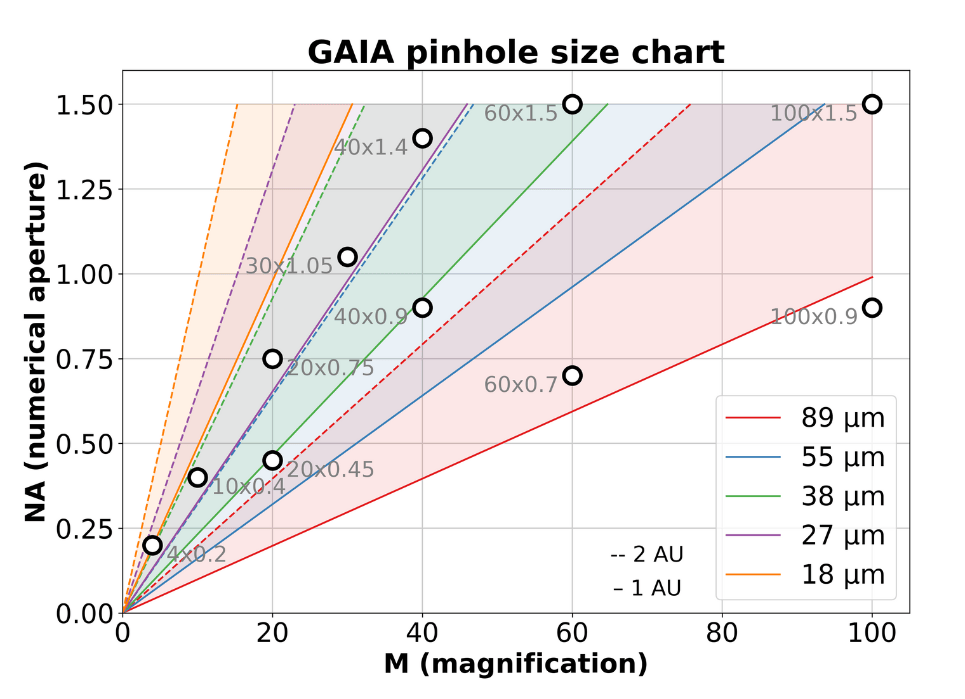
Our flagship GAIA λ has a switchable pinhole, ensuring flexibility, sensitivity and optimal confocality.
Nyquisting every objective and adding fast multicolor imaging over a large FoV gives raise to the most light-efficient super resolution confocal system on the market.
Highlights of GAIA
- Point REscan SR confocal
- SR imaging on large FoV
- Extra deep live cell imaging > 650μm
- Perfected for both VIS and NIR
Discover the way GAIA improves your imaging experience
Video curtesy of Ronald Breedijk, University of Amsterdam
Discover GAIA systems
that meet your needs
GAIA | α | λ |
|---|---|---|
Detector | Camera (sCMOS) | |
Resolution in real time | 120 nm deconvolved, 170 nm raw image | |
Detector sensitivity | Up to 96% QE | |
FOV | FN18: 330×330 μm in super resolution using 40x objective | |
Speed in line scanning | 3 fps at 512 x 512 px, 30 fps in sprint level max at 256 x 256 px | |
Wavelength | VIS+NIR (400-1100 nm) | |
Software | Micromanager, SDK available for integration on request | |
Deconvolution | Microvolution (real time); SVI Hyugens (post processing) | |
Modalities | Super resolution, Widefield, Brightfield | |
Pinhole | Fixed | Switchable |
Emission filter | Single band filter wheel, optional quad band only | Motorized single band filter wheel |
Adaptability | All commercially available bodies | |

See for yourself how we improve your experience
Request your
personalized demo
- Your online demo will be shown in only 45-60 minutes
- Our experts will lead you through our products performance
- We will contact you and together, we pick a day and time

